Polly Beak Deformity Correction
Conveniently located to serve the areas of Beverly Hills and Greater Los Angeles
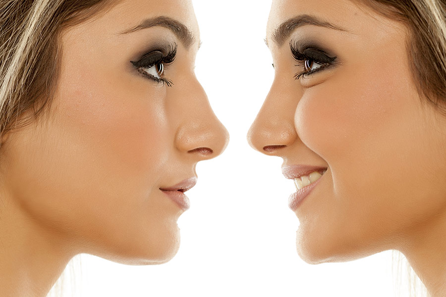
Polly beak, or supratip deformity, occurs when the area above the tip of nose looks too full, or develops a prominent bump after a nose surgery. It’s a common complication that arises after a rhinoplasty, or nose job. Unfortunately, the only way to resolve this is with a corrective surgery known as a revision rhinoplasty. The surgeon will remove or reshape the excess tissue or cartilage to create a nasal tip with more natural-looking projection. This may involve using cartilage grafts to add support to the nasal tip or using suture techniques to reposition the tip. The exact approach will depend on the individual case and the desired outcome.

If you’re looking for expert help with a polly beak deformity, look no further than Michael Omidi, MD, FACS. With almost two decades of experience performing rhinoplasties, he is a highly qualified and experienced plastic surgeon who can help you achieve your desired results. At his Beverly Hills office, he offers a wide range of solutions for both functional and aesthetic issues related to the nose. Whether you’re looking to improve your appearance or simply breathe more easily, he can help. To schedule your personal consultation, call (310) 281-0155 or use our online contact form to get in touch today.
Learn more about the procedures Dr. Omidi performs on his patients by following our blog.
Contents
Why Does Polly Beak Happen?
Polly beak deformity is the most common complication of rhinoplasty and can occur as a result of poorly executed surgery or poor healing. [1] Rather than a smooth, linear nasal profile, patients are left with fullness above the tip of their nose, an area known as the caudal dorsum. This area of tissue creates a curve to the lower nose that causes it to resemble the beak of a parrot, hence the equally unfortunate name for this complication. During rhinoplasties, surgeons trim away, or resect, areas of cartilage in the nose. They often also use sutures to adjust the angling of the various pieces of cartilage that make up the structure of our noses. A recent study that examined the surgical causes of this deformity found that the most common causes of supratip deformity include [2] :
- Over-resection of the nasal tip
- Over-repositioning of the nasal tip
- Inadequate tissue in the nasal tip
- Individual patient characteristics were not fully taken into account during the surgery
Polly beak deformity has been seen in both primary and secondary rhinoplasty patients. In primary rhinoplasty patients, the deformity is typically caused by one or more of the following factors:
- Inadequate tip projection
- An over projected caudal dorsum, or the lower part of the bridge of the nose
- Lower lateral cartilage that is angled incorrectly
In secondary rhinoplasty patients, the deformity is often caused by one or more of these factors:
- Underresected or overresected caudal dorsum
- Overresected mid vault
- Under projected tip (pseudodeformity)
- A combination of these factors
These findings suggest that polly beak fullness can be caused by a variety of factors and that a tailored approach is necessary to address the deformity and achieve the desired results in each individual case.
More About Revision Rhinoplasty
If the top part of the nasal tip still looks too full more than a year after the initial surgery, the only way to fix it is to do another surgery – this is called revision rhinoplasty.
Revision rhinoplasty, also known as secondary rhinoplasty, corrects or improves the results of a previous nose surgery (primary rhinoplasty). Revision rhinoplasty can address a wide range of issues, including:
- Nasal asymmetry
- Difficulty breathing
- Dissatisfaction with the shape or appearance of the nose
- Complications from the initial surgery (such as polly beak deformity, or saddle nose deformity)
Revision rhinoplasty is generally considered to be more complex than primary rhinoplasty, as the surgeon must work with the existing nasal structure, which may have been altered by the previous surgery; this can make it more challenging to achieve the desired results, and a revision surgery may take longer and require more advanced techniques, such as cartilage grafts.
It’s important to choose a qualified and experienced surgeon to perform revision rhinoplasty and the surgeon should have a good understanding of the anatomy and mechanics of the nose.
What are the Benefits of Polly Beak Deformity Correction?
The benefits of correcting a polly beak deformity through revision rhinoplasty include:
Improved Aesthetics
The correction of a polly beak deformity can result in a more natural-looking and aesthetically pleasing nose shape, which can help to boost a person’s self-confidence.
Improved Breathing
In some cases, a polly beak deformity can impede breathing by blocking the nasal passages. Correction of the deformity can restore normal breathing, allowing the patient to breathe more easily.
Improved Functionality
The correction of a polly beak deformity can improve the overall functionality of the nose. It can help to prevent complications, such as nasal obstruction, which can lead to chronic sinusitis.
Improved Self-Esteem
People who are unhappy with the appearance of their nose may experience low self-esteem, social anxiety, and depression. Correcting a polly beak deformity can help to improve their self-esteem and overall well-being.
Because revision rhinoplasty is more complex than primary rhinoplasty, it’s important to choose a qualified and experienced surgeon to perform the procedure to minimize the risk of postoperative complications. To schedule your consultation with Dr. Omidi, call (310) 281-0155 or fill out this form.
Candidates for Surgery
Candidates for revision rhinoplasty should be:
- In good overall health, as the procedure is invasive and requires anesthesia. It’s important that the tissue can heal adequately from the surgery.
- Patients who have chronic sinusitis or are experiencing breathing troubles due to a blocked nasal passage caused by a polly beak deformity.
- Patients who have realistic expectations about the outcome of revision rhinoplasty, as it may not be possible to achieve the same results as primary rhinoplasty, but it can improve the aesthetic and functional aspects of the nose.
- Unhappy with the shape or appearance of their nose.
Revision rhinoplasty should be considered only after the patient has completed the healing process of the primary rhinoplasty and all swelling has resolved. This usually takes about a year.
Personal Consultation
Patients who think they can benefit from a primary or revision surgery to address polly beak deformity, must first start with a consultation. During the consultation appointment, Dr. Omidi will perform a thorough examination of the patient’s nose, including an analysis of the patient’s medical history, the patient’s desired outcome, and the patient’s overall health condition.
Dr. Omidi and his team will also take photographs of the patient’s nose to document the patient’s current condition and to plan the surgical strategy. During the consultation, the patient will have the opportunity to ask questions and discuss any concerns they may have. Dr. Omidi will explain the surgical options, the potential risks and benefits of the procedure, and the recovery process. After the consultation, the patient will have a better understanding of the surgery, the expected results, and will be able to make an informed decision about whether to proceed with the surgery or not.
Preparation
Our team will provide the patient with detailed pre-operative instructions, including information on how to prepare, and what to expect during recovery. Patients should stop smoking and avoid taking certain medications, such as blood thinners, as they can increase the risk of complications. They should also fill any prescription pain medications Dr. Omidi prescribes and plan for a friend or family member to drive them home after surgery.
Furthermore, the patient may need to:
- Get a blood test – The patient may be required to have some blood tests done to check for any medical issues that could affect the surgery.
- Get imaging done – Dr. Omidi may order imaging studies in order to get a more accurate view of the patient’s nose’s internal structure.
Based on the patient’s examination, consultation, and imaging studies, the surgeon will create a surgical plan, which will be discussed with the patient and any questions or concerns will be addressed.
The Procedure
In patients who are undergoing nose surgery for the first time, Dr. Omidi will fix the problem by removing extra skin or cartilage from the lower part of the nasal bridge, adjusting the angle of the tip, or doing a combination of both. The technique will depend on what the primary problem is. Often, the problem is caused by several factors, all of which will need to be addressed during surgery.
If the cartilage on the sides of the nose above the nostrils (the lateral cartilage) is tilted forward, Dr. Omidi can reposition the cartilage and secure them into the correct placement with sutures. In other cases, he may need to trim appropriate amounts of cartilage to restore a smoother line to the profile.
If swelling and scar tissue are contributing to the lingering fullness in the area shortly after the surgery (within 1 to 3 months) Dr. Omidi can use a special tape to help sculpt the healing tissue and resolve the appearance of fullness.
The techniques and methods Dr. Omidi uses during your surgery will be determined by the state of your tissue, your unique characteristics, and more. After the surgery, Dr. Omidi will dress your nose with a nasal splint. This will protect and support your nose during the first part of your recovery. After a brief period of monitoring, you’ll be released from our care, and will need to have a friend or family member drive you home after surgery.
Recovery
Recovery can vary from person to person. In general, patients can expect:
24 Hours After Surgery
The intranasal packing Dr. Omidi applied to the patient’s nose can be removed.
Days After Surgery
Patients may experience some pain and discomfort during the first few days after the surgery. Medications can be prescribed to manage the pain.
Weeks After Surgery
Patients can expect some swelling and bruising around the eyes and nose. This usually subsides within a few weeks to a few months.
Months Following Surgery
Patients are advised to rest and avoid strenuous activities for several weeks after the surgery.
Patients will have several follow-up appointments with Dr. Omidi to monitor the healing process and to ensure that the nose is healing properly. Avoid heavy lifting, bending, or straining, do not sleep on your face, and be very careful to not hit your nose as it could distrupt the healing process and alter your results.
Results
It may take several months to a year for the final results of revision rhinoplasty to be visible. The patient’s nose will continue to change and refine during the healing process.
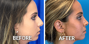
Correcting a polly beak deformity allows patients to enjoy a smoother, more natural-looking nasal profile, and improved self-esteem and overall well-being.
What is the Cost of Polly Beak Deformity Correction in Beverly Hills?
Patients in California can enjoy the results of their polly beak deformity correction in the heart of Beverly Hills at the hands of a skilled, board-certified surgeon. During your consultation, Dr. Omidi will discuss the cost of your procedure. If you feel that the appearance of your nose is the only thing that is holding you back, call (310) 281-0155 today.
References
- Parkes ML, Kanodia R, Machida BK. Revision rhinoplasty. An analysis of aesthetic deformities. Archives of Otolaryngology–Head & Neck Surgery. 1992;118(7):695-701. doi:10.1001/archotol.1992.01880070025005
- Guyuron, Bahman M.D.; DeLuca, Louis M.D.; Lash, Richard M.D.. Supratip Deformity: A Closer Look. Plastic and Reconstructive Surgery 105(3):p 1140-1151, March 2000. https://journals.lww.com/plasreconsurg/Abstract/2000/03000/Supratip_Deformity__A_Closer_Look.49.aspx


